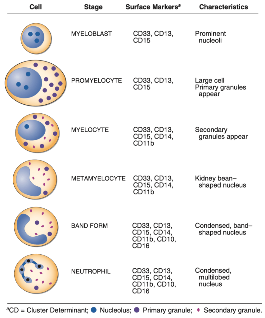
FIGURE 64-2 Stages of neutrophil development shown schematically. Granulocyte colonystimulating factor (G-CSF) and granulocyte-macrophage colony-stimulating factor (GM-CSF) are critical to this process. Identifying cellular characteristics and specific cell-surface markers are listed for each maturational stage.

Döhle Body
FIGURE 64-3 Neutrophil band with Döhle body. The neutrophil with a sausageshaped nucleus in the center of the field is a band form. Döhle bodies are discrete, blue-staining, nongranular areas found in the periphery of the cytoplasm of the neutrophil in infections and other toxic states. They represent aggregates of rough endoplasmic reticulum.
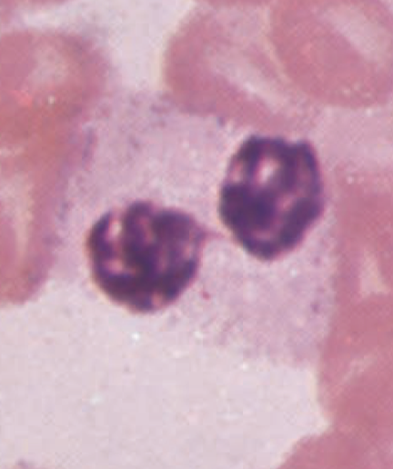
Pelger-Huet anomaly
FIGURE 64-5 Pelger-Hüet anomaly. In this benign disorder, the majority of granulocytes are bilobed. The nucleus frequently has a spectacle-like, or “pince-nez,” configuration.
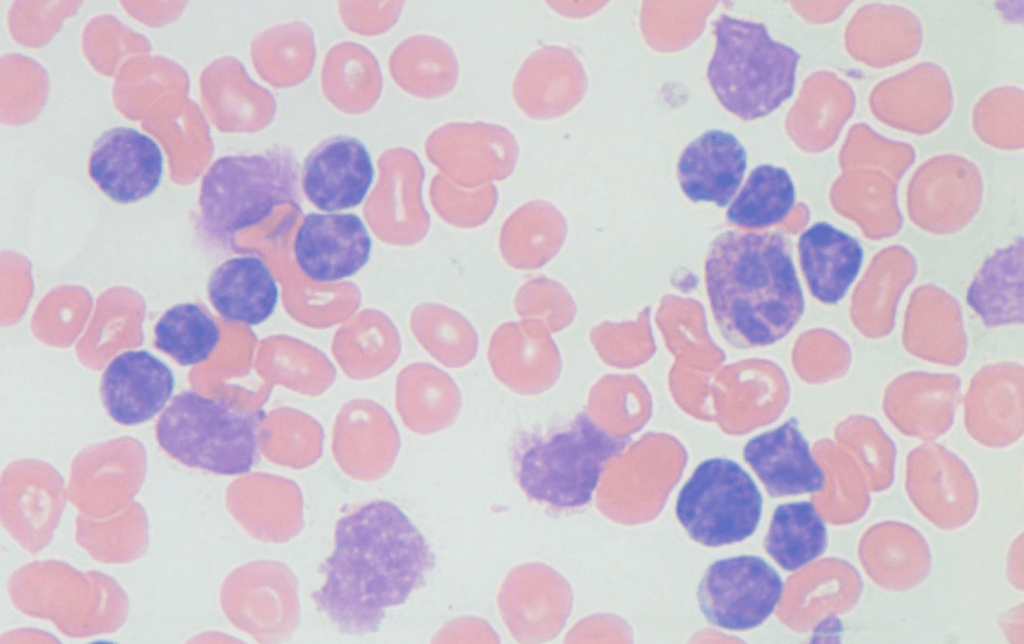
Smudge cells
Usually observed in CLL patients. Mature lymphocytes that rupture easily.
Leave a Reply