T cell activation
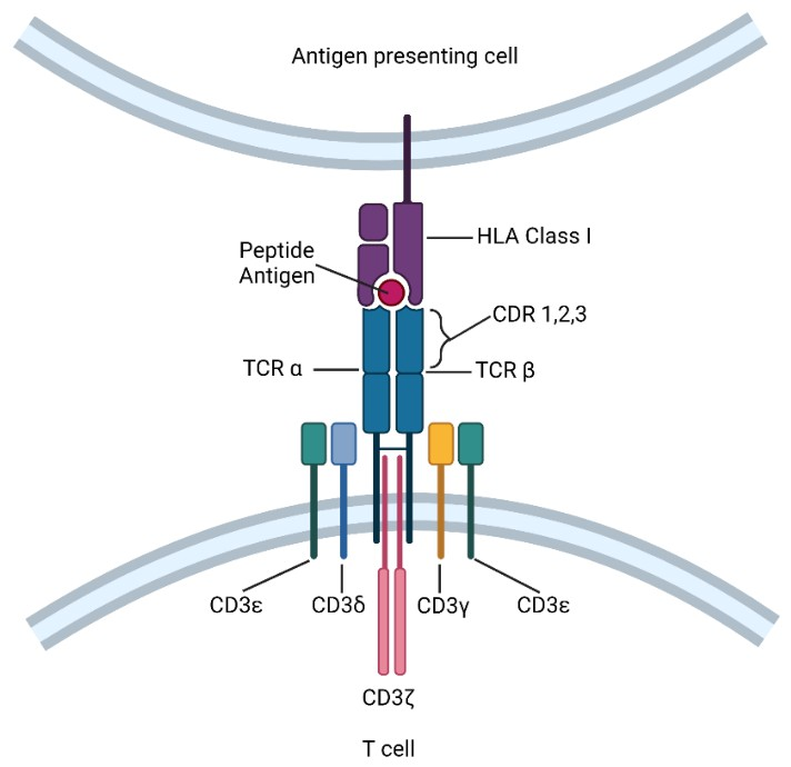

The TCR complex recognizes antigens with MHC molecule
- TCR complex comprises highly polymorphic single α and β-glycoprotein chains
(small T cell population harbours γ and δchains instead) - Rearrangement of α and β-chain -> vast repertoire of T cell clonotypes
Costimulation by CD28 (recognizes B7-1, B7-2)
Activated T cell
In lymphoid tissue
Early after: CTLA4 displaying on the cell surface
- Before being active, CTLA4 is contained within intracellular vesicles in naive T cells
- After activated, trafficking of CTLA-4 containing vesicles is controlled by interaction with LRBA enables
Late after: CTLA4 binds to the B7-1 and B7-2
- CTLA4 and CD28 are very similar, but CTLA4 has greater affinity and avididty than CD 28 for B7 ligands
- 3 mechanisms
- Directly antagonizing CD28, by competing for co-stimulatory ligands
- Thereby preventing their binding to CD28 -> promoting anergy by decreasing the T cell activation state.
Constitutive expression of CTLA4 in Treg
- Endocytosis of B7 ligands
In peripheral tissue
Early after: upregulate PD1 at the mRNA level
Late after: surface expression of PD1
- PD1 binds to its ligands PDL1 and PDL2, thereby T cell exhaustion at sites of infection or when confronted with neoplasms.
CD3 signalling
- Non-polymorphic signalling cahins, by dimeric CD3 γ-δ, ε-δ, ζ-ζ
- intracellular portions of the CD3 <- (inhibitory) phosphorylated by LCK
- Upon activation, CD45 removes inhibitory phosphate on LCK, permitting phosphorylation of ZAP70 -> recrutes LAT and PLCγ
- Calcium release -> activation of GTPase RAS, essential for activated T cell function
Following activation, fates of circulating naive T cells
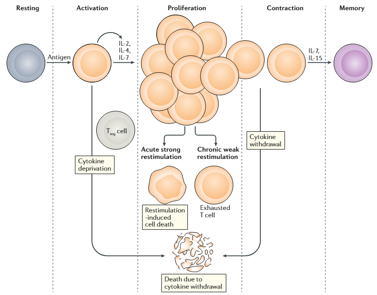
Peripheral T cell fates after antigenic activation. Resting T cells become activated after stimulation by cognate antigen in the context of an antigen-presenting cell and co-stimulatory signals. Activated T cells produce and consume proliferative/survival cytokines, for example, IL-2, IL-4 and IL-7, and begin to expand in number. If CD4+CD25+ regulatory T (Treg) cells are present, they can deprive the cycling T cells of proliferative/survival cytokines, especially IL-2, causing them to undergo apoptosis. Once cells are proliferating rapidly, they have different fates depending on their environment. If they receive acute strong antigenic stimulation, especially if it is encountered repeatedly, the cells will undergo restimulation-induced cell death. By contrast, if they receive chronic weak antigenic stimulation, the cells will survive but become reprogrammed into a specific unresponsive transcriptional state known as ‘T cell exhaustion’. Finally, as the antigen and cytokine stimulation diminishes as the immune response wanes, usually once the pathogen has been cleared, cytokine withdrawal can occur passively to contract the expanded population of antigen-specific T cells. A small fraction of cells will be reprogrammed to enter a ‘memory’ phenotype, and this differentiation step is facilitated by IL-7 and IL-15. Memory T cells will continue to persist in the immune system and form the basis of anamnestic responses. In these regulatory processes, T cell death usually takes the form of apoptosis.
- Apoptosis d/t cytokine withdrawal
Restimulation-induced cell death by acute strong restimulation - Exhaustion by chronic weak restimulation
(e.g., chronic infections and neoplastic processes) - Involved in long-term memory
Immunotherapy
Immune checkpoint blockade
CTLA4
- Very similar to CD28, but has greater affinity and avidity for B7 ligands
- Mechanism of action
- Competing for co-stimulatory ligands (internalization of its ligand)
- Preventing immune conjugate formation (reorganize the cytoskeleton)
- Recruiting inhibitory effectors (e.g. phosphatases like SHP2, PP2A)
- Only for Treg) prime dendritic cells to induce anergy by trans-endocytosis.

CTLA4 blockade in cancer
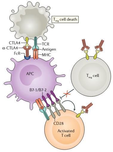
Effects of CTLA4-blocking antibodies. CTLA4-blocking antibodies (α-CTLA4), especially when bound to an Fc receptor (FcR) on an APC, can promote antibody-dependent cellular cytotoxicity (ADCC). CD4+CD25+ regulatory T (Treg) cells express higher amounts of CTLA4 than conventional T cells and are therefore more prone to α-CTLA4-induced ADCC than conventional T cells. In addition, α-CTLA4 can bind to CTLA4 on the surface of the Treg cell and prevent it from counter-regulating the CD28-mediated co-stimulatory pathways that are playing a role in T cell activation. At the same time, α-CTLA4 can also promote T cell responses by blocking CTLA4 on the surface of conventional T cells as they undergo activation.
Mechanism of action
- Boosting effector T cell responses
- Depletes local intratumoral Treg cells through ADCC
Prediction of outcome
- (m/i) ratio of effector T cells to Treg cells
PD1/PDL1
- PD1 exhibits a 20% identity to CTLA4 and 15% to CD28.
Binds the B7 homologoues PDL1 (a.k.a. B7-H1) and PDL2 (a.k.a. B7-DC) - Distinctions from CTLA4 axis
- Spatially, CTLA4 play within lymphoid organs, whereas PD1 play within peripheral tissues.
- Temporally, PD1 acts later in the course of T cell activation and fate determination.
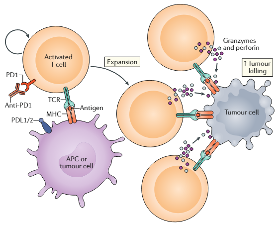
Mechanisms of PD1 axis inhibition. Activated T cells express PD1, which engages with its specific ligand (PDL1 or PDL2) to dampen
activation. Blocking of the PD1 axis through the administration of an anti-PD1/PDL1/PDL2 antibody prevents this inhibitory interaction and unleashes antitumoural T lymphocyte activity by promoting increased T cell activation and
proliferation, by enhancing their effector functions and by supporting the formation of memory cells. Consequently, more T cells bind to tumour antigens presented on tumour cells by MHC molecules via their TCRs. This ultimately leads to the release of cytolytic mediators, such as perforin and granzyme, causing enhanced tumour killing.